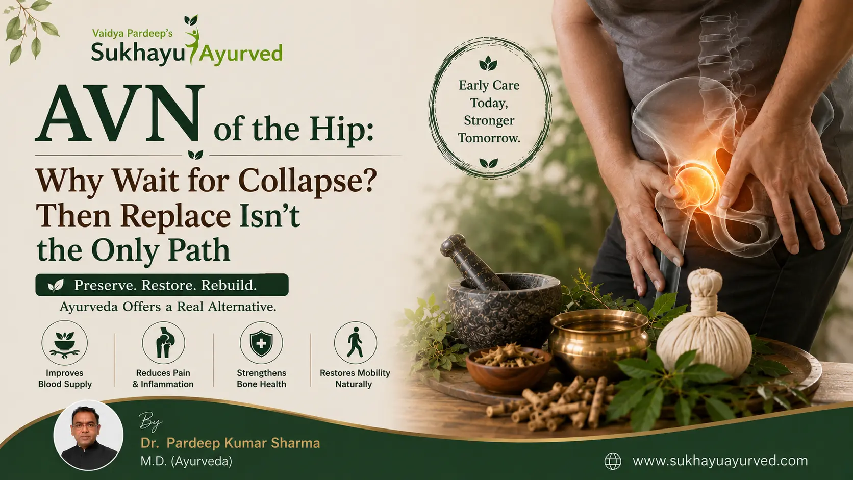
Most patients who reach me with Avascular Necrosis of the hip have already heard the same sentence from at least two different doctors: "This will only get worse. Replacement is just a matter of time." I understand why they say it — in a large number of cases, by the time AVN is caught, it has genuinely progressed too far to manage without surgery. But in the earlier grades, before certain thresholds are crossed, there is real, MRI-documented room to work with the disease instead of waiting it out. This article is about where that room exists, and — just as importantly — where it doesn't.
AVN Is a Blood Supply Problem, Not a Tissue Problem
The first thing I want a patient to understand is this: the necrosis is not the disease. It's the result of the disease. The femoral head dies because its nutritional supply — and its drainage — has been cut off. Once you understand AVN as a vascular obstruction problem rather than a tissue problem, the treatment logic changes. The goal isn't to manage dead bone. It's to restore what's actually failing: the blood supply feeding it.
This is also why AVN so often gets missed for months. Early on, most patients assume the ache is a muscular spasm in the hip. As it progresses, it starts mimicking sciatica — and in my experience, more than 30% of AVN cases aren't correctly diagnosed until they've already reached Grade II. Part of this is the disease's nature. Part of it is historical: before the wave of steroid-induced AVN cases (post-COVID courses of steroids being a major contributor in recent years), orthopedists weren't trained to actively look for it — it was treated as a rare condition, not a differential to rule out routinely.
Is It AVN, or Is It Sciatica?
Because the two conditions share overlapping symptoms — limping, hip-region discomfort, restricted movement — they get confused constantly, and that confusion costs patients time they can't get back. The distinction, in practice, comes down to the character and location of the pain:
| Sciatica | Avascular Necrosis | |
|---|---|---|
| Nature of pain | Neural — sharp, shooting | Muscular, deep-seated |
| Effect of movement | Worsens the pain | Restricted movement, but pain itself is not movement-dependent in the same way |
| Effect of rest | Improves the pain | Does not improve — often worsens at night |
| Radiation | Travels down the leg, past the knee, to the toes | Confined to hip, groin, and knee — rarely radiates below the knee |
If a patient's pain gets worse lying down at night and doesn't extend past the knee, that's a signal to specifically image the hip rather than assume it's a spinal issue.
What We Actually Assess Before Recommending Non-Surgical Management
Not every AVN case is a candidate for Ayurvedic management, and I want to be direct about that, because overclaiming here would put a patient's joint at real risk. We take cases up to Grade IV on the Ficat and Arlet classification — but grade alone isn't the deciding factor. What we look for on MRI is:
- The condition of the bone and the degree of bone marrow edema
- Whether sclerotic or osteoarthritic changes have set in
- Whether there is major structural collapse of the femoral head
If more than roughly 80% of the femoral head is affected, joint space is completely diminished, bone marrow edema is severe, and there is collapse with loss of the femoral head's normal contour — that patient needs surgery. At that point, the joint cannot be managed by Ayurveda or anything else non-surgical, and I tell patients this plainly. Ayurveda is not a delay tactic for an inevitable replacement; it's an alternative path for cases where the joint is still salvageable.
The Ayurvedic Understanding: Vatashonita, Not Just "Vata Imbalance"
I classify AVN as Vatashonita — a classification Acharya Charak himself describes with unusual precision for this condition. Charak Samhita states:
वायुर्विवृद्धो वृद्धेन रक्तेनावारितः पथि । कृत्स्नं सन्दूषयेद्रक्तं तज्ज्ञेयं वातशोणितम् ॥
In essence: aggravated Vata, obstructed in its passage by vitiated Rakta, goes on to vitiate that Rakta completely — and this cycle is Vatashonita. It's a self-reinforcing loop: obstructed blood aggravates Vata, and aggravated Vata further corrupts the blood. This is also one of the very few conditions in classical Ayurvedic texts — alongside Kushtha — where actual tissue necrosis is described as part of the disease process, which tells you Charak understood this as something distinct from ordinary joint disease.
The practical implication: the problem isn't the necrosed tissue itself. It's the obstructed channel supplying and draining that tissue. Treat the obstruction, and you address the cause rather than managing the consequence.
Our Treatment Approach
Because Majja (bone marrow) is the seat of activity in this disease, our primary intervention is Tikta Ksheer Basti, aimed at clearing the Rakta obstruction and pacifying the aggravated Vata simultaneously, rather than treating these as two separate steps.
We also work directly on what I call the kidney axis. In Ayurveda, Vrukka (kidney) is described as the seat of Medas, which is itself the precursor of Asthi Dhatu (bone tissue) — meaning kidney function and bone formation are directly linked in classical understanding. This has an interesting parallel in current research: the kidney's regulation of the Klotho protein and FGF23 is now understood to directly influence osteoclast activity and systemic bone metabolism. We use medicines built around Gokshura and Manjistha to support this axis, alongside Lepana applied at Kati Pradesh (the lower back, considered the root of Vata) to help pacify the aggravated Vata locally.
A typical first schedule runs 16 days of Basti. Most patients report that the midnight pain episodes — one of the most disruptive symptoms of AVN — begin resolving here, along with a reduction in limping. We then repeat an 8-day Basti course after an interval of two to three months, generally for three to four cycles depending on the individual's response.
What Patients Must Do — and Must Not Do
Two restrictions are non-negotiable: complete avoidance of alcohol and smoking, both of which directly compromise vascular health.
For patients managing AVN alongside SLE, RA, or another autoimmune condition — or CKD — I do not tell them to alter their prescribed medication. They continue to follow their treating physician's guidance on those medications; our treatment works alongside that, not against it.
The instruction that surprises most patients is around movement. I ask patients to avoid exercise or excessive movement of the affected joint entirely, which runs against what most people expect for joint problems. Here's why: in AVN, the necrosed patch of the femoral head is rough, not smooth. When patients — often on a physiotherapist's advice — keep moving that joint under load, that rough surface behaves like sandpaper against the smooth cartilage of the acetabulum. I've seen physiotherapy-driven "exercise for pain management" cause acetabular damage that wasn't there to begin with. Rest, in this specific disease, is treatment — not passivity.=
A Case in Point
One of our patients was just 21 years old when he first noticed knee pain during ordinary daily activity. It took roughly six months and visits to multiple specialists before AVN was even considered as a diagnosis. By the time he was diagnosed, his MRI showed subchondral collapse and flattening of the femoral head. Over roughly 1.5 years of treatment with us, his AVN improved from Grade IV to Grade II — a shift documented on follow-up MRI, not just reported symptomatically. His youth was a genuine advantage in how his body responded; not every case, especially in older patients or those with ongoing risk factors like steroid use or alcohol history, will see the same degree of improvement.
The Honest Bottom Line
If your MRI shows major femoral head collapse, near-total joint space loss, and severe bone marrow edema, Ayurveda is not going to reverse that — and no responsible physician should tell you otherwise. But if you're in an earlier grade, with the joint's structure still substantially intact, there is a real, physiologically grounded alternative to "wait and replace." The deciding factor isn't hope. It's what your imaging actually shows.
If you or a family member has been diagnosed with AVN — at any grade — the single most useful next step is getting your existing MRI reviewed against these specific criteria, rather than guessing which category you fall into. [Book a consultation with Dr. Pardeep Sharma] to have your scans assessed and get a direct answer on whether non-surgical management is a realistic option in your case.
This article is for educational purposes and reflects clinical experience in Ayurvedic practice. It is not a substitute for individualized medical evaluation. Ayurvedic management is intended to complement, not replace, conventional diagnosis and care — particularly in advanced-grade AVN, where delayed conventional treatment (including surgery, where indicated) can have serious consequences. Please consult a qualified Ayurvedic physician for assessment specific to your case.